
From DARKNESS…
to LIGHT
Deep Eye Surgery
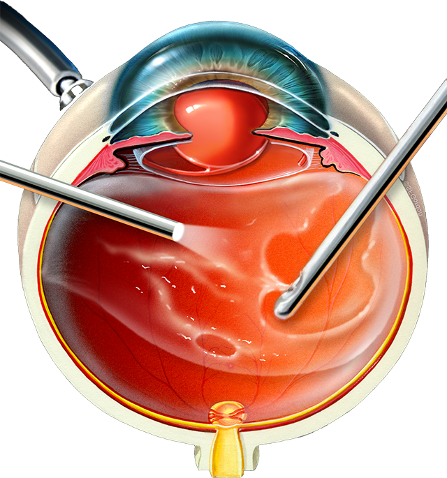
Two years after men landed on the moon in 1969, vitrectomy finally enabled surgeons to reach the back of the human eye. Many of the Foundation’s breakthroughs are enhancements of vitrectomy.
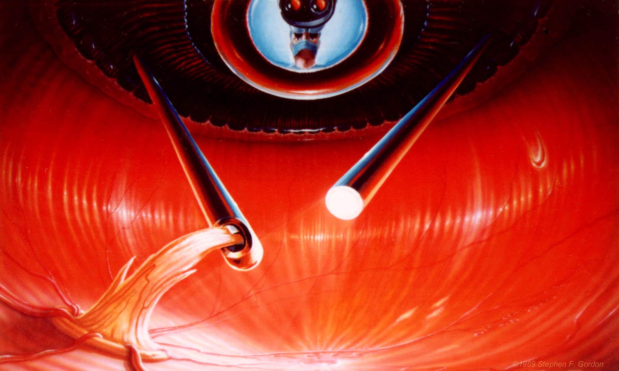
Retinal Detachment Prevention
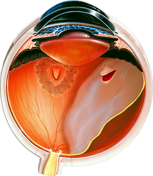
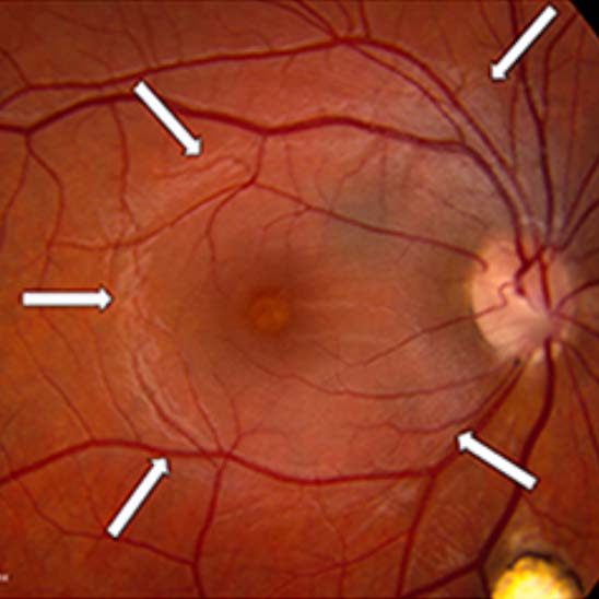
Retinal detachment occurs spontaneously in the aging eye, and it is blinding unless successfully repaired by surgery. The fellow eye is also at risk. Proof of effective preventive treatment eluded researchers for over 50 years despite the introduction of laser treatment for the retina in ~1970.
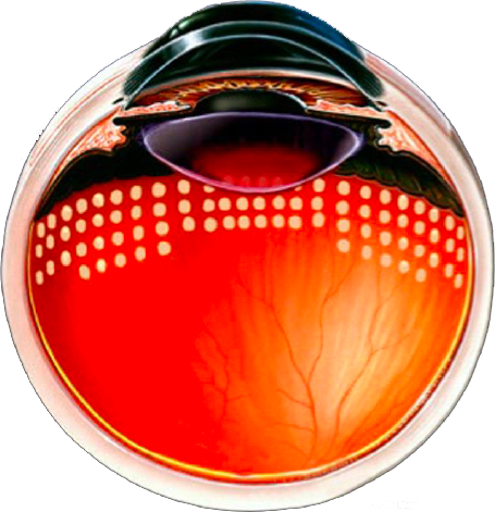
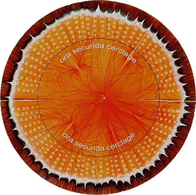
In 2022, HKF published the first strong proof of safe and effective laser treatment for the prevention of retinal detachment in asymptomatic eyes determined to be at high risk. This was accomplished by non-invasive laser “welding” of the peripheral retina in an encircling pattern.
© 1999, 2008 Stephen F. Gordon
Floaters
“Floaters” are opacities in the aging vitreous gel that are common and usually innocuous, allowing 20/20 vision “between floaters.” Occasionally they are extensive, substantially interfering with reading, driving, safely using stairs, or walking across a street. We named this “Degenerative Vitreous Syndrome,” (floaterstories.com).
In 2007, the Foundation produced a surgical video of vitreous opacity removal that won the Buckler Award from the American Society of Retina Specialists. This was the first time any eye society had acknowledged such treatment as safe and effective, hastening treatment for innumerable sufferers who had previously been turned away.
In 2022, the Foundation published “Vitreous Opacity Vitrectomy (VOV): Safest Possible Removal of “Floaters,” reducing the surgical risk of such treatment to ~1%, similar to cataract extraction.
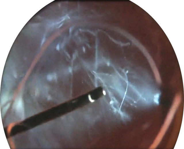
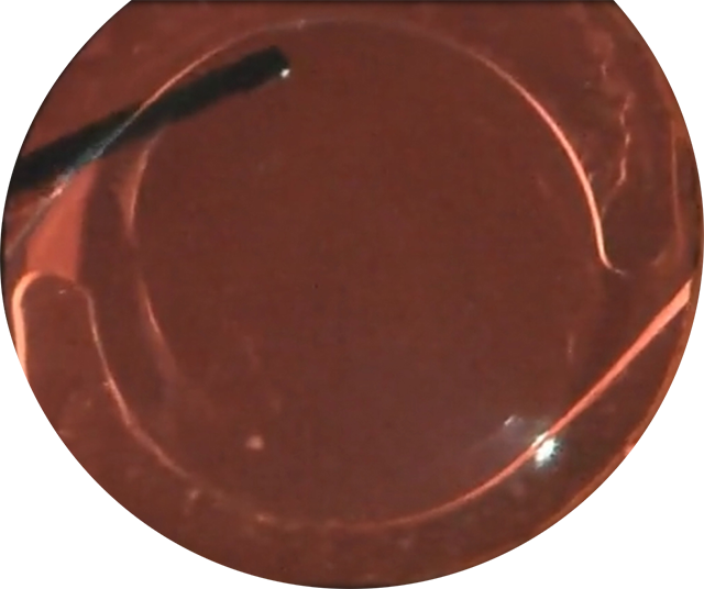
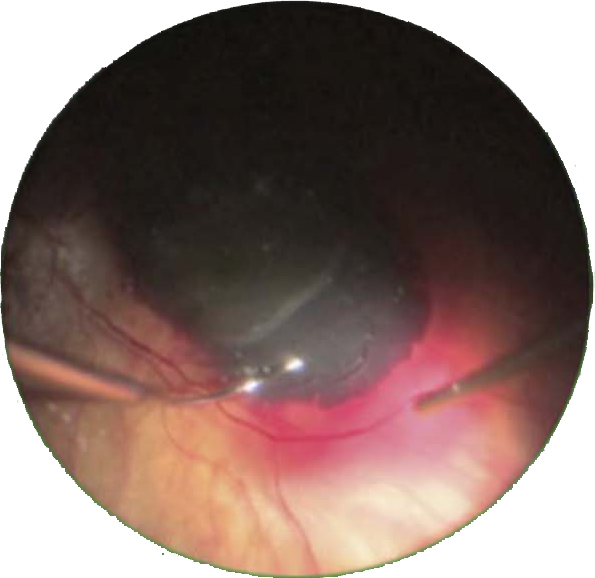
Intraoperative images before and after removal of vitreous gel opacities to restore continuously clear vision.
Internal Limiting Membrane (ILM) Peeling
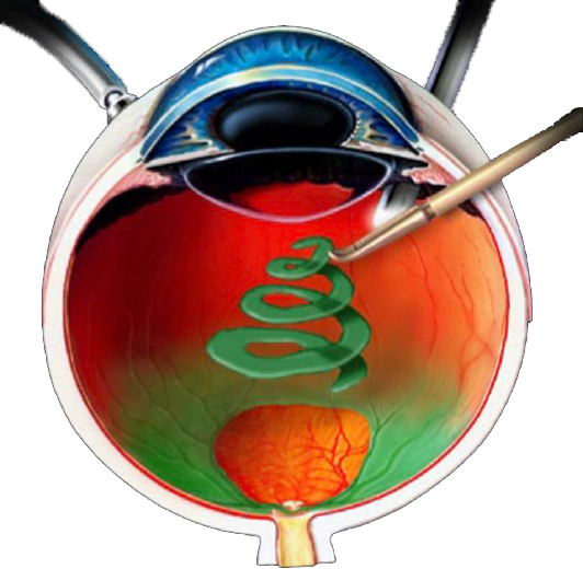
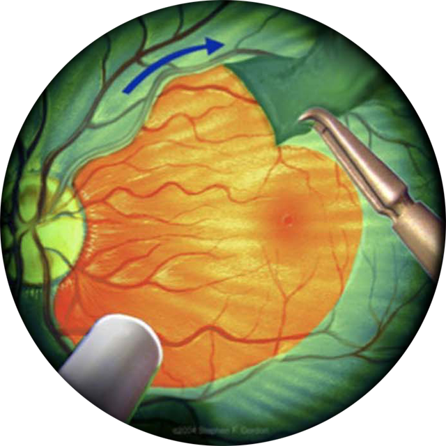
At the annual meeting of the American Academy of ophthalmology in 1990, HKF announced the first ever peeling of the scarred macular surface (ILM), which had previously been considered inviolate. This discovery permanently revolutionized macular surgery.
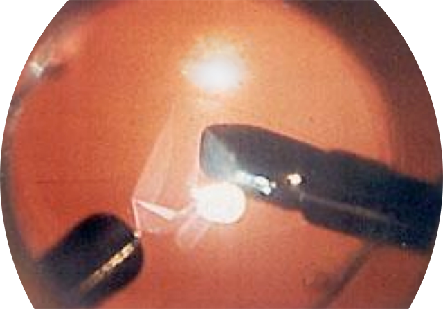
© 2000 Stephen F. Gordon
Macular Degeneration
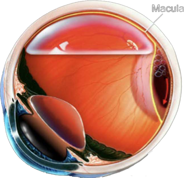
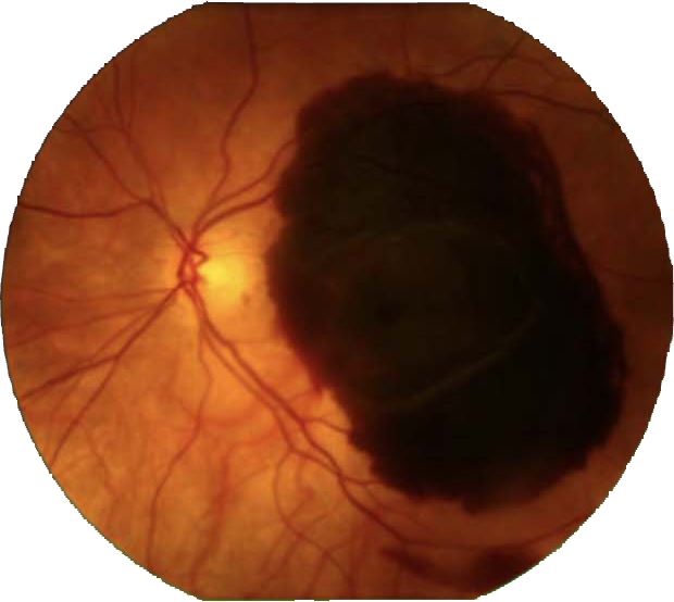
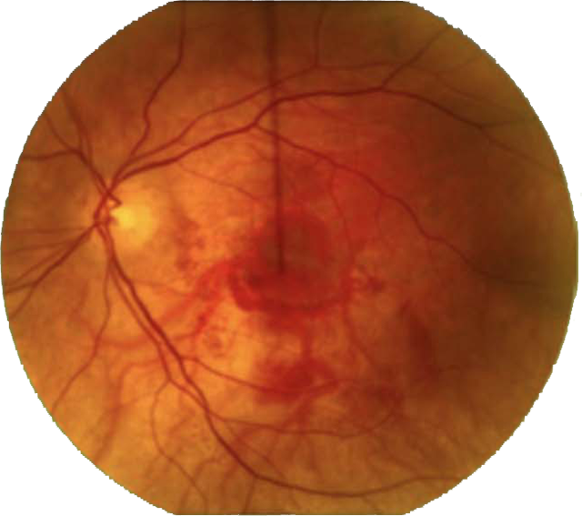
Eyes with wet macular degeneration can suffer sudden large bleeds under the macula, causing immediate loss of vision to the legally blind level. Foundation researchers injected large amount of subretinal fluid with “clot buster” into and around these recent bleeds to mobilize them, and a gas bubble to quickly move them out of the macula. Most eyes regained reading vision postoperatively.
Vitrectomy for Endophthalmitis
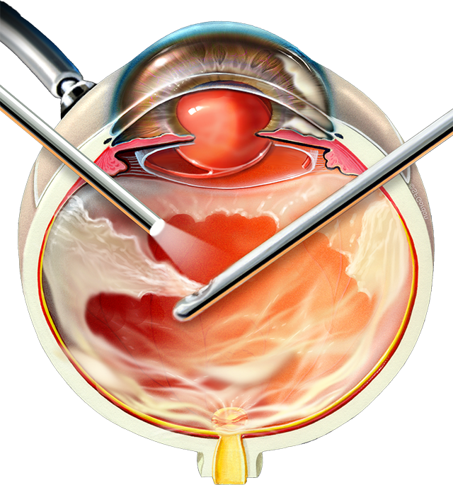
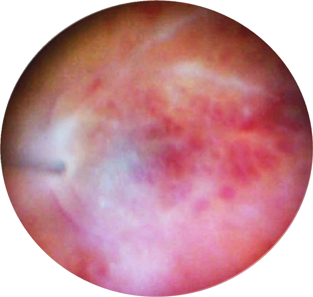
After cataract extraction, most patients quickly achieve excellent vision. Occasionally, an infection inside the eye will appear within days after surgery, profoundly reducing vision. By analyzing 10 years of treatment data. Foundation researchers showed that complete and early vitrectomy surgery for most eyes suffering such infection could restore reading vision in ~80% of cases. This is compared to ~50% using predominantly antibiotic treatment alone, the existent standard treatment.
Congenital Optic Nerve Deformity
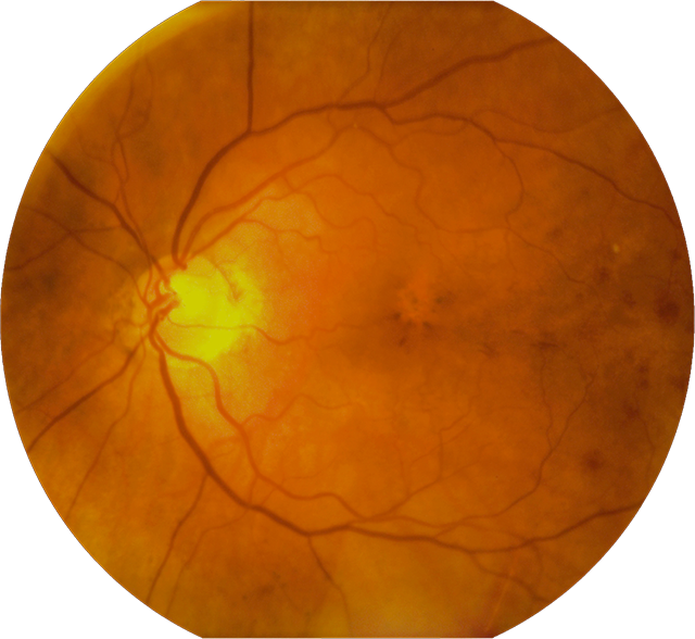
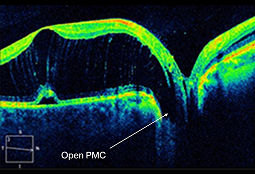
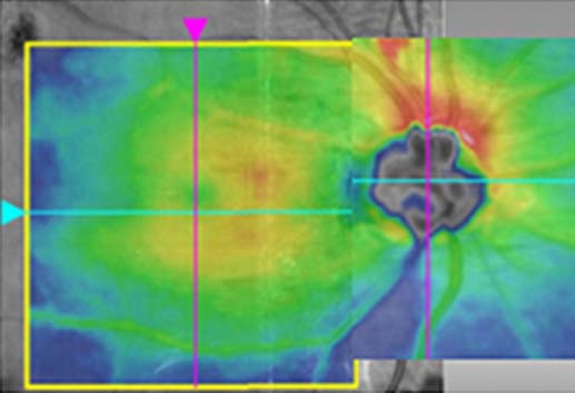
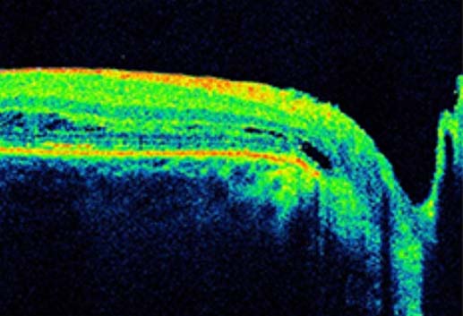
Rarely, the optic nerve has a small defect at birth (optic disc pit, ODP) that allows fluid to spontaneously extend into the adjacent macular, even decades after birth. HKF researchers show that this can be cured by vitrectomy and a long duration gas bubble, without higher risk procedures performed directly on the optic nerve and macula.
Eye Injury
“We walk by faith – not by sight.”
2 Corinthians 5:7
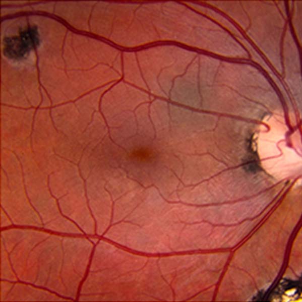
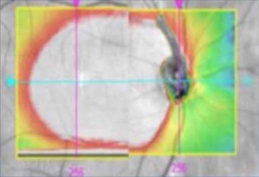
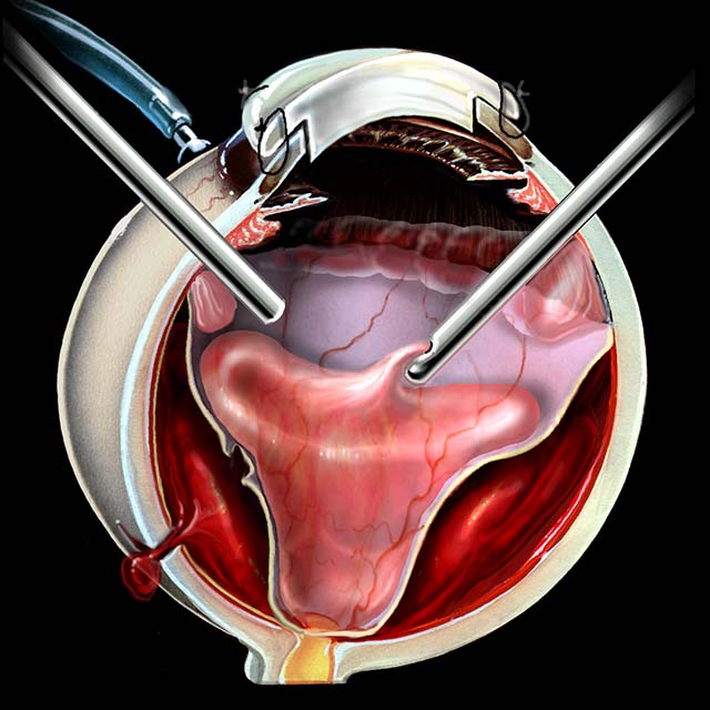
Eye injuries are devastating and effect mostly the young. We showed that many injured eyes could be saved from removal and restored to vision by vitrectomy surgery performed through a plastic lens, temporarily replacing the injured opaque cornea, before finally placing a permanent and clear transplanted cornea.
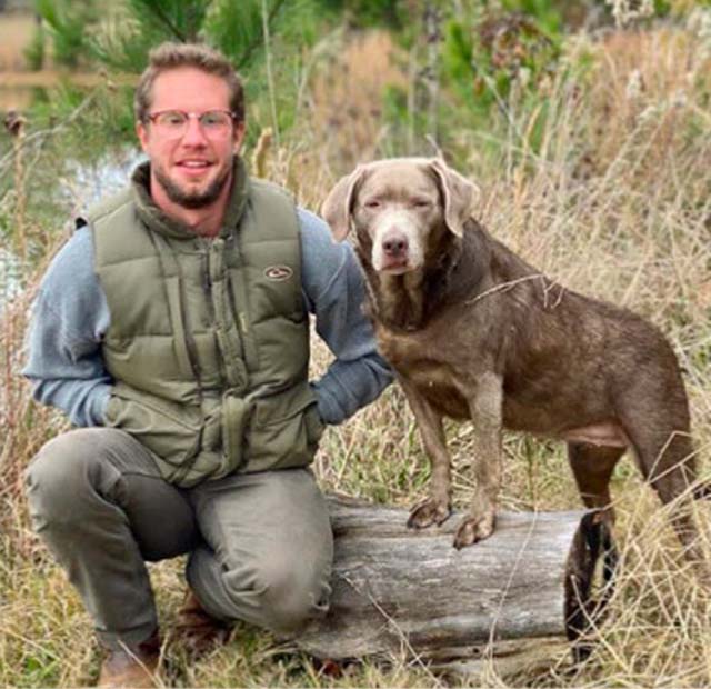
“It is the eye injury surgeon’s responsibility to distinguish reasonable hope from futility in the search for vision. It is an unending, career-long duty.”
Robert Morris, MD
Describing recurrent operations on the “No Light Perception” injured eyes of Cameron Stovall in 2014, ultimately restoring vision in one eye.